


After fertilization, the zygote undergoes a series of divisions and forms a mass of many cells called a morula. Later, a fluid-filled cavity forms within the morula, and the cells are arranged into two groups around it. This structure is called the blastocyst. The cells of the outer layer of the blastocyst are called trophoblasts. When the blastocyst enters the uterus, the trophoblast cells secrete enzymes. These enzymes dissolve the cells of the uterine lining, forming a small cavity into which the blastocyst embeds.
The trophoblast divides rapidly and forms two layers. The outer layer is called the syncytiotrophoblast, while the inner layer is called the cytotrophoblast. The cytotrophoblast cells divide and penetrate the wall of the uterus in the form of finger-like structures called villi, which are surrounded by a layer of syncytiotrophoblast. These villi continue to grow and branch further. They are called chorionic villi.
Blood vessels develop within these villi. The villi become closely associated with the blood vessels in the endometrium, establishing a connection between the mother’s and the baby’s blood circulation without the two blood supplies mixing directly. Later, the villi are embedded in the decidua. Their number increases in the decidua basalis. By the end of the fourth week of pregnancy, the development of the chorionic villi is complete.
By the sixteenth week of pregnancy, the placenta increases in both diameter and thickness. As it develops, the placenta takes the shape of a round, flat disc, with its center thicker than its edges. It weighs about 500 grams. At the time of delivery, the placenta measures about 20 cm in diameter and is approximately 2.5 cm thick at its center.
Umbilical Cord.
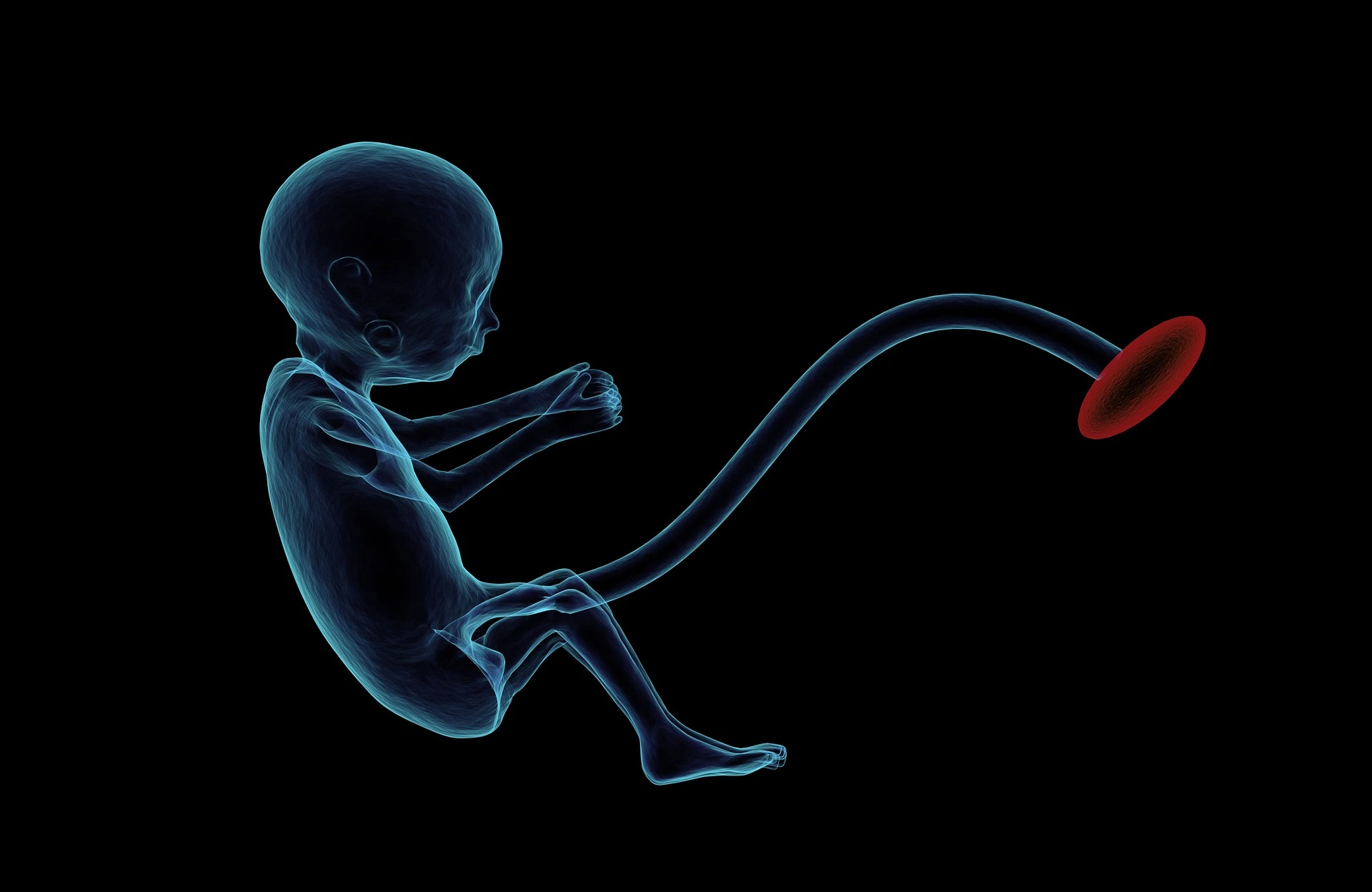
The placenta is attached to the fetus by a cord-like structure called the umbilical cord. It is about 50 centimeters long and 2 centimeters in diameter. It contains the following blood vessels:
- Umbilical Vein – This vein carries oxygenated blood and nutrients from the placenta to the baby.
- Umbilical Arteries – These two arteries are wrapped around the umbilical vein and carry deoxygenated blood and waste products from the baby to the placenta. The umbilical cord is attached to the baby’s abdomen at one end and to the placenta at the other end.
Functions of the placenta.
- Supplies oxygen to the baby and removes carbon dioxide.
- Supplies nutrients to the baby from the mother’s blood.
- Removes waste products from the baby into the mother’s blood.
- The placenta also acts as a barrier, helping to prevent the spread of certain infections from the mother to the baby.
- The placenta secretes various hormones, including:
- Human Chorionic Gonadotropin (hCG)
- Estrogen
- Progesterone
Placenta defects.
- Weight: The placenta normally weighs about 500 grams.
- Location: The placenta is usually located on the anterior or posterior wall of the uterus near the fundus. This is called a normally placed placenta.
- Shape: The placenta is usually disc-shaped. Sometimes, it consists of two or three lobes, and sometimes an accessory lobe is attached to it.
- Accessory Placenta: This is sometimes present at some distance from the main placenta. It is connected to the main placenta by blood vessels.
- Placenta Circumvallata: A white ring is present around the placenta on the fetal side. The folding of the chorionic membrane around the edge of the placenta forms it.
Placental Diseases.
After the death of the baby in the uterus, if the membranes rupture or the duration of labor is prolonged, bacteria can invade the placenta. Due to which various diseases can arise in the placenta.
- Bacteria are difficult to detect in the placenta during the first trimester of pregnancy.
-
Syphilis: In syphilis, the placenta is usually larger and heavier than normal. However, syphilis-causing bacteria are difficult to detect in the placenta during the first trimester of pregnancy.
-
Erythroblastosis Fetalis: In erythroblastosis fetalis, the placenta is usually enlarged, pale, and heavier than normal due to edema and increased blood formation
If the disease is mild, the placenta is less severely affected and may appear yellow due to the deposition of bile pigments. In hydrops fetalis, the placenta is swollen, enlarged, heavier than normal, yellow, and may rupture easily. -
Fibrinoid Degeneration: Almost every placenta has small white areas near its margins. These areas result from fibrinoid degeneration of the trophoblast. In most cases, this is a normal finding and does not interfere with the supply of oxygen and nutrients to the fetus.
-
Intervillous Thrombosis: Sometimes, maternal blood clots form in the intervillous space of the placenta. As a result, blood flow to the surrounding villi is reduced, leading to the formation of red infarct-like areas. If the thrombosis becomes extensive, it may impair the supply of oxygen and nutrients to the fetus.
- Calcium Deposition (Placental Calcification): Small calcium deposits are commonly found on the maternal side of the placenta, especially near term. In most cases, this is a normal finding and does not affect placental function. However, extensive calcification may reduce placental efficiency.
